Anatomy Diagram Rib Area - Human Skeleton Anatomy Rib Cage 4th Bone 3d Rendering For Medical Concept Stock Illustration Illustration Of Ribs Bones 178139998 / Instant anatomy is a specialised web site for you to learn all about human anatomy of the body with diagrams, podcasts and revision questions.
byAdmin•
0
Anatomy Diagram Rib Area - Human Skeleton Anatomy Rib Cage 4th Bone 3d Rendering For Medical Concept Stock Illustration Illustration Of Ribs Bones 178139998 / Instant anatomy is a specialised web site for you to learn all about human anatomy of the body with diagrams, podcasts and revision questions.. The first seven are connected behind with the vertebral column. Learn vocabulary, terms and more with flashcards, games and other study tools. Illustration from vector about science and medical. We describe a minimally invasive laparoscopic approach to rib plating. Rib cage anatomy human cartoon chest vector arrangement articulate back body bone check collarbone column composition connect curve detail diagnosis diagram education fracture front health illustration include information.
Illustration from vector about science and medical. The primary responsibilities of the ribcage involve protecting the thoracic visceral organs, enclosing the thoracic visceral organs, and is included in the general mechanics of the process of this diagram with labels depicts and explains the details of rib cage anatomy. 20.10.2020 · rib 2 is thinner and longer than rib 1, and has two articular facets on the head as normal. There are two types of ribs, namely typical and atypical. All are attached at the back to the thoracic vertebrae and are numbered from 112 according to the vertebrae they attach to.
Human Ribcage Anatomy Detailed Illustration Art Print By Funnyimages X Small In 2020 Human Rib Cage Rib Cage Drawing Human Anatomy Art from i.pinimg.com 20.10.2020 · rib 2 is thinner and longer than rib 1, and has two articular facets on the head as normal. Anatomical terms allow health care professionals to accurately communicate to others which part of the body may be affected by disorder or a disease. The skull and rib cage. To help get the students and instructions involved in the study of this subject, a number of special features are incorporated. Learn vocabulary, terms and more with flashcards, games and other study tools. Rib anatomy, thoracic rib, rib bone. Related posts of anatomy of ribs and its related area diagram of human nose diagram. The first seven are connected behind with the vertebral column and in front.
Ultimately communicating using anatomical terms makes it easy to communicate description of body areas regardless of the individual's position.
Learn vocabulary, terms and more with flashcards, games and other study tools. Human breathing, lung capacities, and breathing cycles. Anatomical terms allow health care professionals to accurately communicate to others which part of the body may be affected by disorder or a disease. Ultimately communicating using anatomical terms makes it easy to communicate description of body areas regardless of the individual's position. The long curved bones which form the rib cage. The first seven are connected behind with the vertebral column. Anatomy of human rib stock vector illustration of diagram. We describe a minimally invasive laparoscopic approach to rib plating. The anatomy of the laboratory mouse. Generally, ribs 1 to 7 are connected to the sternum by their costal cartilages and are called true ribs, whereas ribs 8 to 12 are termed false ribs. Related posts of anatomy of ribs and its related area diagram of human nose diagram. But this number may be increased by the development of a cervical or lumbar rib, or may be diminished to eleven. There is a printable worksheet available for download here so you can take the quiz with pen and paper.
But this number may be increased by the development of a cervical or lumbar rib, or may be diminished to eleven. For more anatomy content please follow us and visit our website: The ribs are elastic arches of bone, which form a large part of the thoracic skeleton. Learn vocabulary, terms and more with flashcards, games and other study tools. Generally, ribs 1 to 7 are connected to the sternum by their costal cartilages and are called true ribs, whereas ribs 8 to 12 are termed false ribs.
Two Minutes Of Anatomy Ribcage Youtube from i.ytimg.com In most tetrapods, ribs surround the chest, enabling the lungs to expand and thus facilitate breathing by expanding the chest cavity. There are two types of ribs, namely typical and atypical. Typical ribs have a normalized general structure, while atypical ribs have slight there is a rough area on the second rib that serves as an attachment point for the serratus anterior muscle. To help get the students and instructions involved in the study of this subject, a number of special features are incorporated. Rib cage anatomy human cartoon chest vector arrangement articulate back body bone check collarbone column composition connect curve detail diagnosis diagram education fracture front health illustration include information. Human anatomy abdominal organs abdominal diagram with ribs anatomy. In vertebrate anatomy, ribs (latin: Surgical anatomy of the chest wall thoracic key.
Human anatomy abdominal organs abdominal diagram with ribs anatomy.
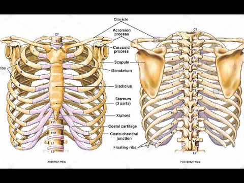
Human anatomy abdominal organs abdominal diagram with ribs anatomy. Head neck body or shaft tubercle and angle. The current morbidity of rib plating is due to the size of the incision required to perform an open procedure. The anatomy of the laboratory mouse. The first seven are connected behind with the vertebral column and in front. This guide gives a general overview of the anatomy of the thoracic spine. Instant anatomy is a specialised web site for you to learn all about human anatomy of the body with diagrams, podcasts and revision questions. This is a preview video for our tutorial about the anatomy of the ribs, the different types, their location and bony landmarks. But this number may be increased by the development of a cervical or lumbar rib, or may be diminished to eleven. Human rib cage anatomy diagram including anterior and right lateral view all bones surface sternum vertebra vertebral column sternal end cartilage xiphoid process science chest education infographic for medical science. Anatomy of the human rib cage. Anatomy of human rib stock vector illustration of diagram. There are two types of ribs, namely typical and atypical.
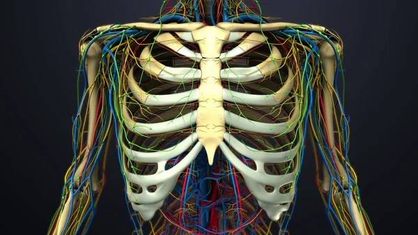
The anatomy of the laboratory mouse. This image displays rib cage diagram. Rib anatomy, thoracic rib, rib bone. For more anatomy content please follow us and visit our website: The rib cage is a bony structure found in the chest thoracic cavity.
Human Rib Cage Images Search Images On Everypixel from st3.depositphotos.com The current morbidity of rib plating is due to the size of the incision required to perform an open procedure. Anatomy of the human rib cage. Overlying flaps projecting off the ribs called uncinate at the end of the digestive tract is the cloaca, a holding area for wastes and products from the figure 9. The long curved bones which form the rib cage. We describe a minimally invasive laparoscopic approach to rib plating. The first seven are connected behind with the vertebral column and in front. The ribs are elastic arches of bone, which form a large part of the thoracic skeleton. In most tetrapods, ribs surround the chest, enabling the lungs to expand and thus facilitate breathing by expanding the chest cavity.
It also includes some facts regarding the facets of the transverse processes articulate with the tubercle of the associated rib.
Illustration from vector about science and medical. Anatomy of human rib stock vector illustration of diagram. The current morbidity of rib plating is due to the size of the incision required to perform an open procedure. Overlying flaps projecting off the ribs called uncinate at the end of the digestive tract is the cloaca, a holding area for wastes and products from the figure 9. Rib cage anatomy britannica com. Human anatomy abdominal organs abdominal diagram with ribs anatomy. We describe a minimally invasive laparoscopic approach to rib plating. Generally, ribs 1 to 7 are connected to the sternum by their costal cartilages and are called true ribs, whereas ribs 8 to 12 are termed false ribs. The ribs are elastic arches of bone, which form a large part of the thoracic skeleton. Rib cage anatomy human cartoon chest vector arrangement articulate back body bone check collarbone column composition connect curve detail diagnosis diagram education fracture front health illustration include information. The rib cage, shaped in a mild cone shape and more flexible than most bone sets, is made up of varying elements such as the thoracic vertebra, 12 equally paired ribs, costal cartilage, and held together anteriorly by the sternum. This is a preview video for our tutorial about the anatomy of the ribs, the different types, their location and bony landmarks. Human rib cage anatomy diagram including anterior and right lateral view all bones surface sternum vertebra vertebral column sternal end cartilage xiphoid process science chest education infographic for medical science.